 3 layers which are all richly supplied with blood vessels:
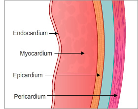
- Epicardium – this is the outer, visceral layer of the pericardium.
- Myocardium – this is the middle, and thickest layer, composed mostly of cardiac muscle cells (myocytes) responsible for cardiac contraction, but also some specialist nerve fibres.
- Endocardium – this is the inner layer of the heart and is a thin layer of endothelium overlaying a thin layer of connective tissue.
The Pericardium is a double layered sac that surrounds the heart.
Structure of Cardiac Muscle
Like skeletal muscle, cardiac muscle fibres are striated, with a single nucleus. They differ in that they are branched and connected by intercalated discs, which allow for very rapid impulse transmission. During embryonic development around 1% of muscle cells become autorhythmic (ie they have the ability to spontaneously depolarise and generate action potentials) and become part of the conduction system, which means they can generate impulses outwith central nervous system control.
Smooth muscle is involuntarily (i.e. you don”t have to think about it!) and is also found in the gastro-intestinal and respiratory tracts.
Smooth muscle
Blood vessels contain a layer of smooth muscle (thicker in arteries than in veins and capillaries) which controls the dilation or constriction of the vessels – important to the regulation of perfusion and blood pressure.
Skeletal muscle
Skeletal muscle fibres are multinucleated and appear striated.
Cardiac and skeletal muscle cells contain compartments called sarcomeres which make the muscle look striated. The sarcomeres shorten during contraction.
Page last reviewed: 19 May 2020