At the hospital

Jimmy and his wife arrive at the Emergency Department of the thrombectomy hub. There is a local protocol for the management of patient’s arriving with a suspected stroke which should be followed. Protocols provide a clear written description of appropriate action required under specific circumstances.

  1. Taken into area defined locally for hyperacute assessments (perhaps Resus in ED, or a specific bay, or in some hospitals into the scanning dept
  2. Met by doctor and nurse
  3. Rapid assessment to confirm diagnosis of probable stroke – determine deficits with the National Institutes of Health Stroke Scale (NIHSS) – all medical and nursing staff involved in hyperacute stroke care should complete training in this
  4. Assess for eligibility for thrombolysis and thrombectomy (onset time, severity warrants hyperacute treatment)
  5. Insert iv cannula to allow injection of contrast for CTA, and administration of thrombolysis if appropriate
  6. Arrange immediate CT and CTA (if eligible for both thrombolysis and thrombectomy)
  7. If imaging ready take patient through, don’t wait for porters!
How do you elicit an accurate onset time? Answer True or False to each of the questions below:

Learning points

Module aim:

The loss of the ability to communicate effectively after a stroke has a significant impact on many aspects of daily life. This module will examine three key areas: how to support communication, right hemisphere communication difficulties and living with a communication difficulty.

Learning points:

Supported communication
  • Understand what supported communication is and how to use it with someone who has aphasia.
  • Learn about the level of disability that arises for an individual who requires support with communication.
  • Be able to recognise a wide range of resources and strategies that may be used to support communication, depending on an individual’s specific communication support needs.
Right hemisphere communication difficulties
  • Understand the difficulties associated with right hemisphere stroke.
  • Learn about the appropriate strategies and resources to use with someone who has right hemisphere communication difficulties.
  • Be aware of the social, psychological and emotional consequences that occur due to a right hemisphere communication difficulty and how they affect individuals and their family/carers.
Living with a communication difficulty
  • Understand how the communication disorders, aphasia and dysarthria, impact on everyday life for individuals and their family/carers.
  • Gain knowledge about the resources, support networks and aids available to assist individuals to live with a communication difficulty.
  • Appreciate the longer term issues of living with a communication difficulty such as accessing employment and leisure activities and making psychological adjustments.

Time is Brain

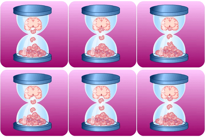
Egg timer with brains animating from top section to bottom

Thrombolysis and/or thrombectomy treatment reduces the risk of residual disability 3-6 months after stroke in appropriately selected patients, even taking into account some early risks. Although the use of thrombolysis is associated with an increased risk of bleeding and death in the first week, there is no overall increased risk of death at 3-6 months.

The chances of a full or nearly full recovery from stroke are much greater the earlier treatment is given. Early treatment within 4.5 hours for thrombolysis, and 6 hours for thrombectomy depends on:

  1. early recognition of stroke symptoms and taking appropriate action
  2. streamlining the flow through the in-patient pathway

We aim to deliver thrombolysis within 30 minutes of arrival at hospital (door to needle time (DTN)) in many cases, and almost always within 60 minutes

The main steps on the pathways to delivery of these hyperacute treatments are shown below, along with some timescales.

These times will depend a lot on the geography of the hospital. The aim is to give the bolus of thrombolysis as soon as possible and certainly within 60 minutes from arrival at hospital.

Introduction – Module Authors

Introduction - Speech bubble

Module lead

Sheena Borthwick, Specialist Speech and Language Therapist, NHS Lothian

Group members

Eleni Pateraki, Principal Clinical Psychologist, NHS Greater Glasgow & Clyde

Emma Coutts, Speech & Language Therapist, NHS Grampian

Gillian Capriotti, Speech & Language Therapist, AHP Stroke Consultant, NHS Greater Glasgow & Clyde

Katrina McCormick, Clinical eLearning Project Manager, RGN, Chest Heart & Stroke Scotland

Natalie Buchanan, Advanced Speech & Language Therapist, Fife Rehabilitation Service, NHS Fife

Sandra Hewitt, Speech & Language Therapist, NHS Highland

Serena Battistoni, eLearning Interactive Content Developer, Chest Heart & Stroke Scotland

Reviewer

Alice Hobbs, Speech & Language Therapist, Stockport NHS Foundation Trust


This module was originally developed by:

Module authors

Module lead: Gillian Currie, Stroke Education Facilitator, Lothian and Borders, CHSS

Group members: Niall Broomfield, Consultant Clinical Psychologist NHS Greater Glasgow & Clyde
Gillian Capriotti, Senior Speech and Language Therapist, NHS Greater Glasgow & Clyde
Joanne Curtis, Specialist Speech and Language Therapist, NHS Lanarkshire
Craig Forman, Charge Nurse, NHS Lanarkshire
Lorna Lowdon, Speech and Language Therapist, NHS Ayrshire & Arran
Jayne McKerrow, VSS Regional Manager (West Scotland), Chest Heart & Stroke Scotland

Critical readers: Sheena Borthwick, Clinical Specialist Speech and Language Therapist, NHS Lothian
Charlie Chung, Clinical Specialist Occupational Therapist in Stroke, NHS Fife
Prof Martin Dennis, Professor of Stroke Medicine, University of Edinburgh
Sharon McGrory, Communication Training Team Manager, Chest Heart & Stroke Scotland
Professor Catherine Mackenzie, University of Strathclyde
Gill Murray, Specialist Physiotherapist, NHS Lothian

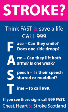
The FAST test

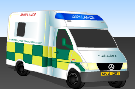
Calling 999 will ensure Jimmy receives urgent medical attention as he may be having a stroke and might benefit from treatment with thrombolysis and/or thrombectomy.

The paramedic uses the FAST tool to help identify patients with stroke. The paramedics also pre-alert the receiving hospital to expect a case of suspected stroke.

For more detailed information visit: FAST Tool: Chest, Heart & Stroke Scotland

How to do the FAST test

Facial movements:

  • Ask patient to show teeth, is there an unequal smile or grimace?
  • Note which side does not move well

Arm movements:

  • Lift the patient’s arms together to 90º if sitting, 45º if supine and ask them to hold the position for 5 seconds before letting go, does one arm drift down or fall rapidly?
  • If one arm drifts down or falls, note whether it is the patient’s left or right

Speech:

  • Listen for new disturbance of speech
  • Listen for slurred speech, get patient to say “British Constitution or Baby Hippopotamus”
  • Listen for word-finding difficulties with hesitations. This can be confirmed by asking the patient to name objects that may be nearby such as a cup, chair, table, keys, pen
  • Check with any person who knows the patient, is this normal for them?

Time to ring 999

13: Communication following stroke

Introduction

On completion of this module you should have a critical understanding of communication problems which are commonly seen after stroke and the impact these can have on the individual.

The answers to all the test questions are contained within the module. This information may be provided in the ‘Additional Information’ boxes on some of the pages.


Case 1: Jimmy

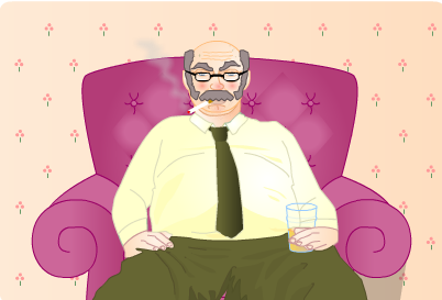
Jimmy is a 58 year man. He is presently unemployed and smokes heavily. He has a family history of ischaemic heart disease and hypertension.

Jimmy Govan sitting in a chair with a cigarette and beer

Jimmy has driven to his local shopping centre with his wife. Whist at the centre he falls over and experiences right-sided face, arm and leg weakness, and speech problems.

Conclusion

Conclusion - Completed jigsaw

Having completed this module and should now:

  • Have knowledge of visual problems following a stroke
  • Have knowledge of assessment processes and screening tools for visual problems after a stroke
  • Recognise the importance of the identification and differentiation of different visual problems which have similar presentations, but may have differing origins and treatment requirements.
  • Understand the impairment and functional limitations which may occur with different visual problems
  • Have knowledge of the referral pathways for visual assessment and interventions after a stroke
  • Have an understanding of the treatment approaches and strategies which may help people with visual problems after a stroke
  • Have knowledge of the professionals and services who can help people with visual problems after a stroke

‘Time is Brain’

Egg timer with brains animating from top section to bottom

Every minute counts following a stroke as millions of brain cells (neurones) die as a result of the interrupted supply of oxygen and glucose.

When an artery blocks, initially the nerve cells stop working which causes the symptoms of the stroke. However, unless blood flow is restored the brain cells start to die, causing permanent damage. This process starts within minutes, but can continue for hours.

Patients admitted with stroke within four and a half hours of definite onset of symptoms, who are considered suitable, should be treated with either intravenous alteplase by infusion over one hour usually 0.9mg/kg (up to maximum 90mg) or an intravenous bolus of Tenecteplase 0.25mg/kg. See Additional Information for more details on dosage.

In patients where a CT angiogram shows that a large artery is blocked a thrombectomy may be indicated if it can be done within six hours of a known time of symptoms onset, or where advanced brain imaging suggests it might be effective.

The delay from stroke onset to treatment with thrombolysis and/or thrombectomy should be minimised. Systems should be optimised to allow the earliest possible delivery of treatment within the defined time window.

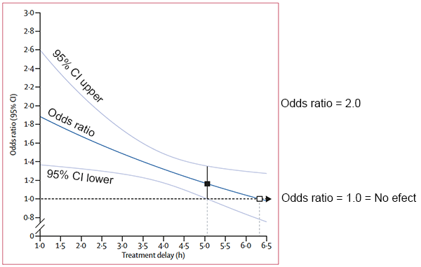
The chances of a full or nearly full recovery from stroke are much greater the earlier hyperacute treatment is given. This graph shows that the effectiveness of thrombolysis is greatest when given early and reduced with increasing delay between stroke onset and treatment. It may not work at all after 270 minutes (4.5 hours).

Alteplase treatment timing graph
Effect of timing of alteplase treatment on the chances of a good outcome (modified Rankin scale (mRs) = 0 (no symptoms) or 1 (minor symptoms)

The odds ratio is the odds (or chance) of a good outcome with treatment divided by the odds (or chance) of a good outcome without treatment. An odds ratio of more than one indicates that it will improve patient outcomes. Therefore an odds ratio of two would indicate a doubling of the chance of a good outcome. The 95% confidence intervals (CI) indicate how precise the estimate of effectiveness is. If the confidence intervals do not overlap the one then we are more confident that the treatment is effective.

“The white box shows the point at which the estimated treatment effect crosses 1.0 the line of no effect.

The black box shows the point at which the lower 95% CI for the estimated treatment effect first crosses 1.0 the line of no effect.”

Lancet 2014; 384: 1929-35

See the full article:

Thrombolysis within 3 hours produced 10 additional good outcomes /100 treated, if the delay was 3-4.5 hours only about  5 good outcomes /100 were achieved –  so 5 fewer patients have a good outcome per 100 treated:

A similar relationship between increasing delay and reducing effectiveness is seen for thrombectomy. Thrombectomy within 6 hours produced 40 patients with less disability /100 treated but for every additional hour of delay to thrombectomy – 7 fewer patients have a good outcome per 100 treated.

Advanced brain imaging (see Additional Information) can be used to identify the small proportion of patients who either wake from sleep with symptoms, or who have had symptoms for more than 4.5 or 6 hours, who might still benefit from thrombolysis or thrombectomy respectively.

For more detailed information:

What is a thrombectomy?

A thrombectomy:

  • Involves putting a thin tube (a catheter) into an artery, usually in the groin
  • Passing it up into the brain, under X Ray guidance, near the blockage
  • Using a small wire net (stentriever) (see video 1 below) or suction (video 2) to remove the clot causing the blockage
  • Removing the clot through the leg artery – A bit like “Dyno-Rod”

Video 1: Irish Heart Foundation: Thrombectomy Animation (YouTube) – showing the use of a stentriever.

Video 2: Mount Sinai Health System: Animation of Thrombectomy Using Aspiration (YouTube) – showing use of a aspiration catheter.